Widefield Microscopy
This automated upright widefield microscope from ZEISS is the right choice for imaging standard glass slides containing fixed- or live-cell samples. Color and monochrome cameras are available for Brightfield/Phase/DIC and fluorescence imaging, respectively. A ten-position filter turret gives the Imager.Z2(M) large flexibility in the fluorophores it can image without the need to stop and swap filter sets.
ZEISS' ZEN blue software package controls the microscope and offers multi-color imaging in conjunction with tile scanning, Z-stacking and time-series acquisition. Advanced tiling functionality allows users to easily incorporate a focus map that accurately follows the changing plane of sectioned tissue; capturing Z-stacks in order to account for focal disparities is no longer necessary, and file size on large tile images is greatly reduced.
The ApoTome.2 optical sectioning device offers confocal-like image quality through the use of structured illumination. Compatible with all lenses and filter sets, ApoTome.2 improves fluorescence image quality by filtering out out-of-focus light to reveal detail in just the focal planes of interest.
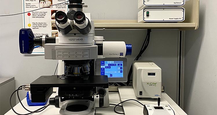
Zeiss AxioImager.Z2(M) for fixed- or live-cell samples
This automated upright widefield microscope from ZEISS is the right choice for imaging standard glass slides containing fixed samples. Color and monochrome cameras are available for Brightfield/Phase/DIC and fluorescence imaging, respectively. A ten-position filter turret gives the Imager.Z2 large flexibility in the fluorophores it can image without the need to stop and swap filter sets.
ZEISS' ZEN blue software package controls the microscope and offers multi-color imaging in conjunction with basic tile scanning, Z-stacking, and time-series acquisition.
The ApoTome.2 optical sectioning device offers confocal-like image quality through the use of structured illumination. Compatible with all lenses and filter sets, ApoTome.2 improves fluorescence image quality by filtering out out-of-focus light to reveal detail in just the focal planes of interest.
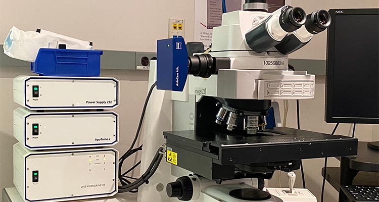
Zeiss AxioImager.Z2 for fixed- or live-cell samples
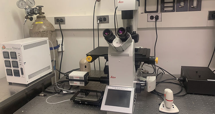
This flagship inverted microscope from Leica features a motorized XY stage for tiled fields and Navigator software module for automated tile acquisition; a TokaiHit stage-top incubation system and time-lapse module for long-term live-cell imaging; and a SpectraX four-LED illumination system capable of fast, multi-color widefield fluorescence imaging. The system is equipped with both color and monochrome cameras for Brightfield/Phase/DIC and fluorescence imaging, respectively. Operated via Leica's LAS X software suite, the DMi8 easily images live- and fixed-cell samples in Petri dishes, well plates, chamber slides, and standard glass slides. Fast widefield FRET imaging (CFP/YFP) can also be configured. Injection of drugs is possible via openings in the stage-top incubation system.
This automated inverted microscope has multiple features to enable long-term time lapse imaging experiments: a LiveCell enclosed chamber that permits temperature, CO2, and humidity control; adapters for well plates, dishes and slides; and a Ludl motorized stage and focus motor that can be used to mark multiple sites in xyz. Images are captured with a Q Imaging Retiga 2000R cooled CCD camera; 1600 X 1200 pixels, 7.4µm X 7.4µm per pixel, sensitivity is high and linear through ~550nm and then decreases linearly between 600nm to ~900nm. Exposure to light is controlled by Uniblitz shutters. The system is controlled by MetaMorph software. An on-site tissue culture incubator is available to house cells prior to imaging. Typical uses for this system include cell tracking, neurite growth, cell motility, wound healing, and Fura-2 imaging.
At left, Olympus Live Cell System. At right, images of the same specimen at increasing magnification taken by Crystal Pristell
The Axioplan 2ie is outfitted for studies of samples under polarized light. Standard glass slides can be examined under linear and circularly-polarized light, and basic brightfield is available as well. The AxioCam MRc camera coupled with AxioVision software make this a simple-to-use polarized light imaging system.
Axioplan2 microscope is equipped with optics for brightfield (left), plane polarized (center) and circularly polarized (right) light microscopy. Lamb hoof stained with picosirius red by Virginia Gillespie.
This manually-controlled Olympus stereoscope is ideal for imaging whole organs or even entire embryos stained with brightfield or fluorescent markers. The system features a dual-chip Olympus DP80 camera that allows researchers to easily switch between a color sensor for brightfield imaging and a monochrome sensor for higher-sensitivity fluorescence imaging. Larger, tiled fields can be built manually by the user. A 10x zoom factor coupled with optional 0.63x, 1.0x and 2.0x front lenses permits a very large range of magnifications for imaging. The MVX10 is controlled by Olympus' cellSens software suite.
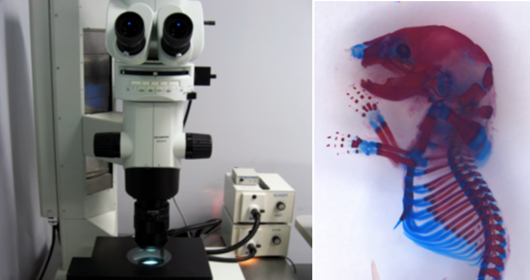
At left, Olympus Macroscope. At right, skeletal prep from an E16.5 mouse stained with Alizarin Red (bone) an Alcian Blue (cartilage) by Jianyun Yan.