Electron Microscopy
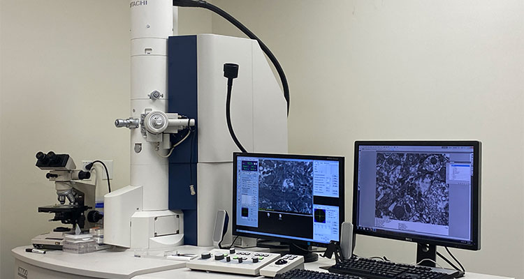
The HT7700 is a state-of-the-art transmission electron microscope. It has Hitachi’s “Dual-Mode” objective lens that helps to achieve superior low magnification, high-contrast as well as high magnification, high-resolution imaging. All image acquisition is digital and there are options that can be used to readily examine and align serial sections. It is equipped with an AMT XR41-B 2K-by-2K digital camera. The HT7700 improved upon the features of the H-7500 and is the 4th generation of PC based TEM’s that uses only a digital camera to obtain images.
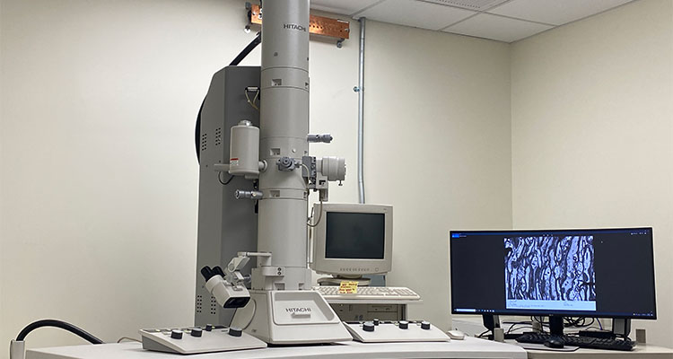
The Hitachi HT7500 transmission electron microscope is equipped with a phosphor screen and an AMT NanoSprint12: 12 Megapixel CMOS TEM digital camera. The system is designed for observation and evaluation of specimens using the electron beam maximum acceleration of 120 KV. Wide fields of magnifications are available both within the low and high magnification functions. Focal distance is selectively usable between high-resolution and high-contrast applications by means of a double-gap objective lens. The main PC integrally controls the TEM main unit and digital AMT camera. A secondary PC is attached to allow user to track and assemble serial images via image software. The H-7500 was Hitachi’s first pc-based TEM sold in the USA. The H-7500 has a phosphor screen and can take pictures with film or use a digital camera.
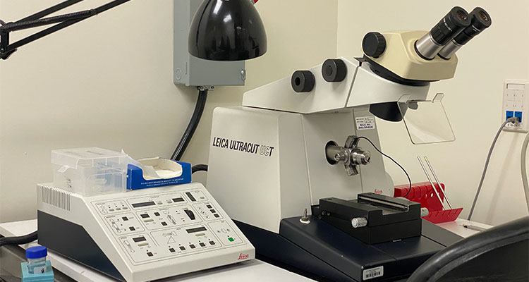
Sectioning capabilities for the CoRE EMs is provided by the UCT and UC7 ultramicrotomes. The systems are capable of section thicknesses down to 60 nanometers for ultrathin slicing; normal sectioning is performed at 80-90nm for ultrathin slices and 0.5-1.0 micron for semi-thin slices. Epon- or lowicryl-embedded samples are sliced according to the sample type, and sections are then mounted on mesh or slot grids as appropriate. Training is not typically provided on this instrument but is possible with strict approval or extensive prior experience.
TEM Services
- Fixative/ Chemical preparation
- Perfusion of large (NHP) to small animal models
- Vibratome sectioning
- Embedding
- For morphological/ultrastructural preservation studies- Embed 812 "Epon"/ Durcapan embedding- (tissue, cultured cells, suspended cells, cell fractions, viruses)
- For Immunohistochemical studies- Freeze substitution resin embedding-Lowicryl HM20 Resin- (tissue, suspended cells)
- High Contrast embedding for FIB-SEM/ 3View
- Ultramicrotome Sectioning
- Semi-thick (Toluidine Blue Stain on slides- 0.5µm- 1µm thick)
- Ultrathin- 2D (minimum 60nm-90nm)
- Serial Sectioning for 3D volumetric studies
- Contrast Counterstaining (Uranyl Acetate and Lead Citrate)
- Pre-screening for CRYO TEM samples
- Negative staining (e.g., viral suspension, exosomes)
- Deionizer & carbon coater on site
- Pre-embed Immunogold labeling
- Post-embed Immunogold labeling