Two-Photon Microscopy
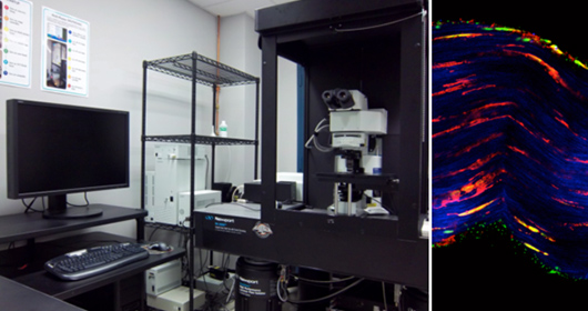
The Olympus FluoView FV1000MPE is a multi-photon laser scanning microscope that allows fluorescence imaging deep within specimens. It has a Coherent Chameleon Vision II laser, tunable from 680nm to 1080nm, as well as a 473nm laser diode for single photon excitation. It has four external (non-descan) detectors, as well as three confocal PMTs and one transmitted light detector. It is capable of timelapse and FRET imaging experiments. The motorized stage contains adapters for different dishes, slides, and small animals to accommodate a wide range of experimental setups, and is also capable of automated montage acquisition and stitching to generate high resolution images of large areas. Dipping lenses allow imaging directly into specimens in media.
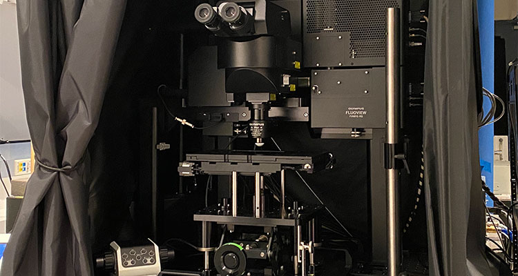
The Olympus FluoView FVMPE-RS multiphoton microscope allows fluorescence imaging at higher depths within the specimen. The upright microscope provides space for large samples and a high degree of motorization. With its ultra-stable arch-like structure, the gantry microscope system offers a high degree of flexibility to suit different samples. This is ideal for in vivo observation requiring maximum space. To facilitate simultaneous two-color excitation, it is equipped with Spectra-Physics Insight X3 tunable ultrafast lasers, having a dual output (I) tunable laser output from 690nm to 1300 nm (II) Fixed laser line at 1045. In addition to the high NA 10X and 25 X objectives, the microscopeis equipped with a computer-controlled correction collar system that can automatically adjust to compensate for spherical aberration during deep observation of thick samples. There are 4 non-descanned detectors on the microscope, 2 PMTs and 2 GAAsP detectors. The motorized stage can be used to image from slides, dishes and small animals. The Core also has an E-Z Anesthesia machine to help intra-vital imaging experiments.
In addition, the microscope is equipped with an Inner Focus Articulating Nosepiece that can be mounted on the microscope to provide more degrees of freedom using a moveable arm while also facilitating rapid axial focusing with no mechanical movement. The software aids in acquiring large mosaic tiles to image large regions. Together with all these features, the microscope can be used for high-sensitivity, high-resolution deep tissue imaging, intra-vital imaging, optogenetics, electrophysiology, label-free imaging (SHG, THG), etc.