Confocal Microscopy
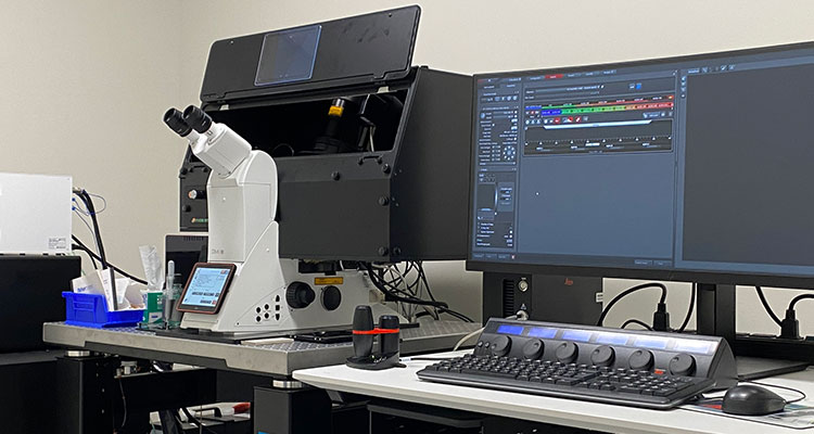
The Leica Stellaris 8 microscope is a confocal imaging system designed to maximize the number of probes and photon collection sensitivity for a wide range of applications, including fixed and live samples. The Stellaris 8 is equipped with a number of technologies that allow it to achieve this versatility and sensitivity. A White Light Laser (WLL) can pump 8 individually tunable and simultaneous laser lines, and it is tunable from 440 nm to 790 nm in 1nm steps. The system features an additional UV405 diode laser bringing the total usable lasers to 9 simultaneously. The system is equipped with 4 spectral PowerHyD detectors having a detection range from 405nm to 850 nm, a photon sensitivity of up to 58%, and a dynamic range up to 180MCounts/s. The spectral capabilities of the Stellaris 8 are augmented by the TauSense technology. TauSense allows real-time separation of fluorophores based on their lifetime properties. It achieves this by computing the distribution of the average arrival time (AAT) of photons. The user is then able to separate fluorophores or remove auto-fluorescence based on these lifetime properties. Tiling and imaging multi-ROI samples is easily achieved in the Navigator interface, and an Okolab incubation box controls temperature, humidity, and CO2.
The Stellaris 8 system is also equipped with Lightning Super-Resolution, a deconvolution-based strategy (Richardson-Lucy algorithm coupled to a statistical decision mask that computes SNR in voxels of data), that can improve resolution to 120 nm in the lateral dimension and 200 nm in the axial dimension. The algorithm can run in a global or adaptive setting depending on the user selection, while preserving the sum of photons in the volume and raw confocal data and runs in near-real-time on up 5 simultaneous detection channels without the need for calibration.
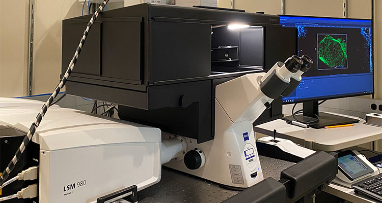
The Zeiss LSM 980 is equipped with Airyscan 2 for super-resolution imaging. The microscope is optimized for simultaneous spectral detection of multiple weak labels, from 405 to 730nm excitation, with high efficiency. The availability of 405, 445, 488, 514, 561, 594, 639 and 730 nm laser lines and an ensemble of 2 multi-alkaline PMTs, 32-channel spectral GaAsP, and 2 near-IR GaAsP detectors can accommodate multitude of fluorophore configurations. The software can also automatically perform experiments like FRAP, FCS, spectral unmixing, linear unmixing with fingerprinting, and large area 3D tiling.
Airyscan 2 is an area detector with 32 circularly arranged detection elements, each of which is acting as a small pinhole contributing to super-resolution information. The highest achievable resolution of the system is 120nm. The Airyscan 2 can also operate in two different multiplex modes to combine super-resolution with high acquisition speed by using parallel pixel readouts. The high sensitivity of the detector combined with the low excitation laser power required and the fast acquisition speeds, makes the Airyscan 2 ideal for sensitive live-cell time series. In addition, it can perform ultrafast time series of single slices, tiling of large areas, or volumetric time-lapse imaging.
The system features a full incubation suite for temperature, humidity, O2 and CO2 control, making it ideal for live-cell studies as well as for inducing hypoxia. The microscope is equipped with a piezo Z-drive, definite focus, and the AI sample finder to automate focus adjustment and identify the relevant imaging regions on the carrier, thus reducing the time to set up the imaging experiment.
The microscope is also equipped with an LED illuminator (Colibri 385, 475, 555, 630nm), a quad cube (DAPI/GFP/Cy3/Cy5), and a widefield camera for fast widefield experiments that can take advantage of the AI finder, incubation, and autofocus hardware.
Leica TCS SP8 with five lasers
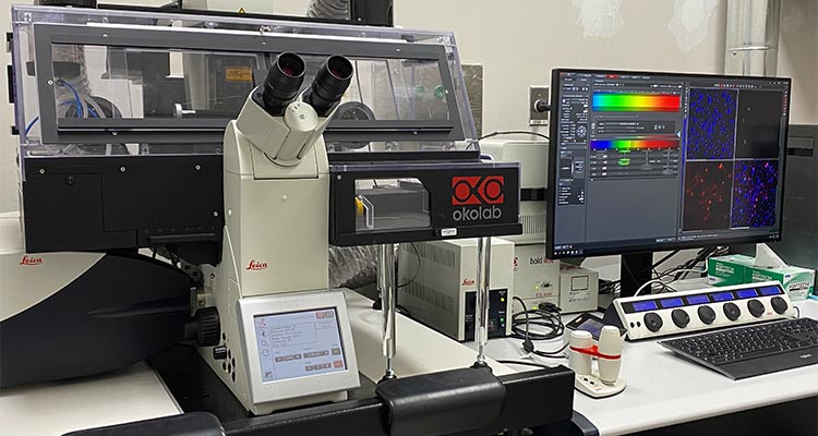
The Leica TCS SP8 sits on an inverted Leica DMi8 microscope stand and has 5 lasers: a UV Diode (405nm), an Argon laser (458nm, 488nm, 514nm), a 561nm, a 594nm and a 633nm. Its spectrophotometer scan head is comprised of 1 PMT and 3 HyD detectors (more sensitive for weaker signals) to detect five different tunable emission wavelengths, as well as a transmitted light detector for the addition of DIC imaging. The motorized stage and “Navigator” software package allow the user to map large sample areas at high resolution in addition to imaging multiple, non-contiguous single positions. Full OkoLab incubation is available as well for live-cell experiments. Hardware is controlled by Leica LAS X software. Leica’s “Lightning” mode is also available, allowing for 40 frames-per-second multi-color imaging at 512x512 pixels, down to 120nm lateral resolution
|
Objectives |
1) HC PlanApo 10x/0.4 "A" |
Requires 0.17mm coverglass |
air/dry |
|
|
2) HC PlanApo 20x/0.75 "C" |
Requires 0.17mm coverglass |
air/dry |
|
|
3) HC PlanApo 40x/1.3 "D" |
Requires 0.17mm coverglass |
oil immersion |
|
|
4) HC PlanApo 63x/1.4 "E" |
Requires 0.17mm coverglass |
oil immersion |
|
|
5) HC PlanApo 100x/1.4 "D" |
Requires 0.17mm coverglass |
oil immersion |
|
|
6) (none) |
|
|
|
|
|
|
|
|
Excitation |
405nm |
|
|
|
|
458nm |
|
|
|
|
488nm |
|
|
|
|
514nm |
|
|
|
|
561nm |
|
|
|
|
594nm |
|
|
|
|
633nm |
|
|
|
|
|
|
|
|
Detectors |
3x HyD |
Frequency Range: 10-1800Hz |
|
|
|
1x PMT |
Detection Range: 400-800nm |
|
|
|
1x T-PMT (for transmitted light) |
|
|
|
|
|
|
|
|
Dichroics |
458/514/561 |
|
|
|
|
488/561/633 |
|
|
|
|
458/514 |
|
|
|
|
488/594 |
|
|
|
|
RT 15/85 |
|
|
|
|
|
|
|
|
Compatible with: |
standard slides (76x26mm) |
|
|
|
|
standard well plates |
|
|
|
|
|
|
|
|
Accessories |
Partial environmental controls |
|
|
|
|
Lightning mode - 40FPS @ 512x512; 120nm XY resolution |
|
|
Long a workhorse of the Microscopy CoRE, the Leica SP5 is a seven-laser line inverted imaging system with many capabilities. The Leica SP5 features six detectors for simultaneous imaging (4 PMT, 1 HyD, 1 T-PMT); a high-speed resonance scanner for up to 25 frames-per-second at 512x512 resolution; a full temperature- and CO2-controlled incubation chamber; the ability to perform multiple types of FRET imaging; and the ability to perform spectral scanning in order to accurately separate overlapping emission spectra. A motorized XY stage easily adapts to different sizes of dishes, chamber slides and standard glass slides. The LAS AF software suite makes setup and image acquisition easy while giving users the opportunity to expand on their imaging capabilities as their needs evolve.
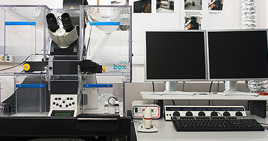
The Leica SP5 DMI microscope with environmental control
Technical Specifications
|
Laser Lines |
Detectors |
Emission Filters |
Dichroics |
Scan Mode |
Bit Depth |
Resolution |
|
405 Diode |
4 PMTs |
Tunable |
RT 30/70 |
Line |
8, 12, 16 |
max: 8192 x 8192 |
|
Argon (458, 476, 488, 514) |
1 Trans PMT |
DD 488/561 |
Frame |
min: 16 x 16 |
||
|
DPSS 561 |
1 HyD |
RSP 500 |
ROI |
|||
|
HeNe 633 |
Substrate |
Reson-ance |
||||
|
TD 488/561/633 |
Spectral decon-volution |
|||||
|
DD 458/514 |
Time lapse |
|||||
|
Montage |
||||||
|
FRAP |
||||||
|
FRET – AB, SE |
||||||
|
Live Data |
Objectives
|
Magnification |
Immersion |
NA |
|
10x |
Air |
0.4 |
|
20x |
Air |
0.7 |
|
40x |
Oil |
1.25 |
|
63x |
Oil |
1.4 |
|
63x |
Glycerin |
1.3 |
|
100x |
Oil |
1.4 |
|
20x* |
Multi |
0.7 |
* available on request
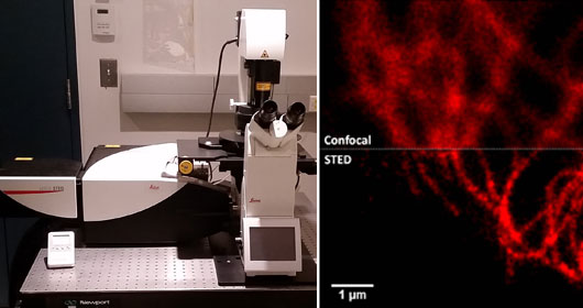
Building on the powerful Leica SP8 confocal scanhead, this super-resolution system features three separate additional lasers for Stimulated Emission Depletion (STED) across a range of florophores. The STED 3x setup provides XY resolution down to 50 nanometers and improved Point Spread Functions for optimal resolution with any sample.
In addition to five standard laser lines and five detectors (2 PMT, 3 HyD), the STED system has a pulsed tunable White Light Laser that will excite fluorophores from 470-670nm. The WLL gives users the ability to precisely maximize excitation efficiency of one fluorophore at a time in multiplexed samples while also enabling the use of advanced imaging techniques such as FLIM, which requires a pulsed laser. The pulsed WLL coupled with the pulsed STED 775nm laser line even allows for FLIM STED, a super-resolution technique that permits separation of emission signals based on temporal data. "Tau STED,” the latest addition to this system, aids researchers with time-correlated photon counting, giving users the unique ability to separate fluorescence signals based on time-of-detection and the actual number of photons collected. Availability of Phasor Plot analysis tool with the module enables effective FLIM and FRET-FLIM studies.
The LAS X software suite from Leica simplifies setup for even the most complex setups.
Technical Specifications
|
Laser Lines (nm) |
Detectors |
Scan Mode |
Bit Depth |
Resolution |
|
Diode Laser 405 nm |
2 PMT |
Line |
8 |
max: 8192 x 8192 |
|
Argon Laser (458, 476, 488, 514) |
3 HyD |
Frame |
12 |
min: 16 x 16 |
|
White Light Laser (WLL) 470-670nm |
T-PMT (transmitted light) |
STED |
|
|
|
|
|
ROI |
|
|
|
|
|
Spectral decon-volution |
|
|
|
|
|
Time lapse |
|
|
|
|
|
Montage |
|
|
Objectives
|
Magnification |
Immersion |
NA |
|
10x |
Air |
0.4 |
|
20x |
Air |
0.75 |
|
40x* |
Oil |
1.3 |
|
63x |
Oil |
1.4 |
|
93x ** |
Glycerin |
1.3 |
|
100x ** |
Oil |
1.4 |
* available on request
** suitable for STED imaging
Harnessing the power of a 32-channel, honeycomb-shaped array detector, the LSM880 with Airyscan offers super-resolution microscopy without the complications of restricted sample preparation or fluorophore choice - if your sample is compatible with a standard confocal microscope, it is compatible with the super-resolution Airyscan detector as well. Resolution improvements up to 140nm in XY and 400nm in Z are possible in combination with imaging speeds of up to 13 frames per second at 512x512 resolution.
The LSM 880 with Airyscan is built on the inverted Axio Observer.Z1 microscope and features a full incubation suite for temperature and CO2 control, making it ideal for live-cell studies. Multispectral imaging is also possible with the LSM880's native 32-channel Quasar detector and two additional PMT detectors: the entire spectrum is scanned at once to quickly build a database of relative fluorophore signal contribution from control samples, and the system can then easily separate emission signals that exhibit high spectral overlap during live imaging of multiplexed samples. DIC imaging is also possible through the use of a Transmitted PMT detector.
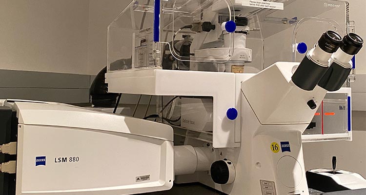
Zeiss LSM880 with Airyscan and environmental control
The LSM880 with Airyscan runs on ZEISS' ZEN black software platform. The software incorporates Tile Scan, Z-stack, Time Lapse and Bleaching functions for multidimensional experiments.
Technical Specifications
|
Laser Lines (nm) |
Detectors |
Scan Mode |
Bit Depth |
|
Diode Laser 405 nm |
32 channel GaAsP |
line |
8 |
|
Argon Laser 458/488/514nm |
T-PMT (transmitted light) |
frame |
12 |
|
DPSS laser 561nm |
2-PMT |
|
16 |
|
HeNe laser 594/633 nm |
Airyscan detector |
|
|
|
Acousto-optical tunable filter |
|
|
|
Objectives
|
Magnification |
Immersion |
N.A. |
Working dist (mm) |
|
10X |
air |
0.45 |
2.0 |
|
20X |
air |
0.8 |
0.55 |
|
40X |
oil |
1.4 |
0.2 |
|
40X* |
water |
1.2 |
0.28 |
|
63X |
oil |
1.4 |
0.17 |
|
100x |
oil |
1.46 |
0.1 |
* available upon request
The LSM780 system sits on an upright Axio Examiner microscope stand with a fixed stage. It has seven laser lines (see below) and is equipped with two fluorescence PMTs, one PMT for transmitted light, and a highly sensitive GaAsP detector that can capture 32 channels simultaneously. Up to 13 images can be captured per second at 512 X 512 pixels. Linear scanning makes this microscope suitable for a variety of quantitative image measurements such as fluorescence correlative spectroscopy. Hardware is controlled by Zeiss Zen Black software, which also incorporates some image processing options. Microscope is enclosed by a C02, 02, humidity and temperature-controlled chamber for imaging living samples
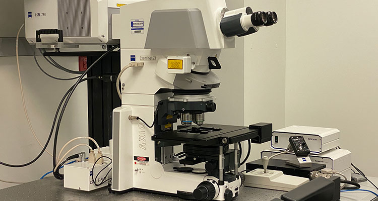
Zeiss LSM780 with a darkened environmental control box
Technical Specifications
|
Laser Lines (nm) |
Detectors |
Scan Mode |
Bit Depth |
|
Diode Laser 405 nm |
32 channel GaAsP |
line |
8 |
|
Argon Laser 458/488/514nm |
T-PMT (transmitted light) |
frame |
12 |
|
DPSS laser 561nm |
2-PMT |
|
16 |
|
HeNe laser 594/633 nm |
Airyscan detector |
|
|
|
Acousto-optical tunable filter |
|
|
|
Objectives
|
Magnification |
Immersion |
N.A. |
Working dist (mm) |
|
10X |
air |
0.45 |
2.0 |
|
20X |
air |
0.8 |
0.55 |
|
40X |
oil |
1.4 |
0.2 |
|
40X |
water |
1.2 |
0.28 |
|
63X* |
oil |
1.4 |
0.17 |
|
100x* |
oil |
1.46 |
0.1 |
* available on request
The CoRE's solution when optical sectioning and high-speed imaging are jointly required is the Yokogawa Spinning Disk Confocal. Built on the inverted ZEISS Axio Observer.Z1 frame and controlled by MetaMorph, the "SD" system is a powerful tool for observing fast live-cell phenomena where cells may not grow in a monolayer but where individual cell responses are needed, such as during drug treatments involving real-time injections. Illumination is provided by the Vortran VersaLase laser launch, with six excitation lasers (405/445/488/514/561/642nm) for multi-color imaging. A motorized stage provides opportunities to image multiple areas of a sample automatically over time. Stage-top incubation for short-term maintenance of cell viability is also available; features include a heated stage insert with cover, humidity and CO2 control, and an objective heater ring. Imaging is provided by the Yokogawa CSU-X1 spinning disk module and two Hamamatsu ImagEM EMCCD cameras for better low-light, rapid imaging; up to two fluorescent channels can be imaged simultaneously. On-site tissue culture equipment is also available, should researchers require it. This system also doubles as a conventional widefield microscope when coupled with the PCO.edge on the left camera port
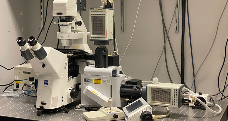
Yokogawa spinning disk confocal microscope
Objectives
|
Magnification |
Immersion |
NA |
|
20x |
Air |
0.4 |
|
40x |
Air |
0.6 |
|
40x |
Oil |
1.3 |
|
63x |
Oil |
1.4 |
|
100x |
Oil |
1.4 |